Congenital Anomalies
Renal Agenesis
Renal agenesis occurs when the urteric bud fails to develop and as a consequence eliminating the induction of metanephric mesoderm.
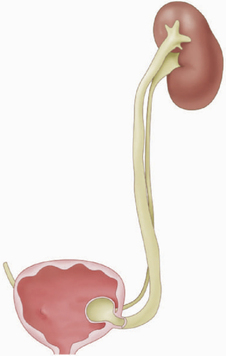
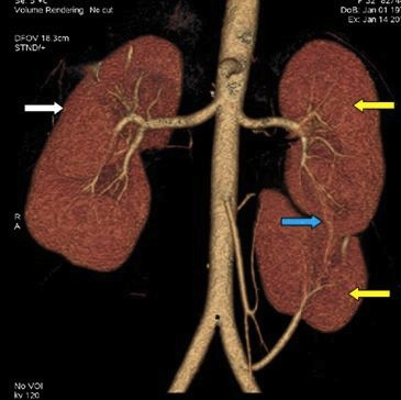
a. Unilateral Renal Agenesis: It is a relatively common anomaly which occurs predominantly in males. In this condition, there is a hypertrophy of the remaining kidney, because it tries to compensate for the missing kidney. As a result this condition usually remains unnoticed because it is asymptomatic and compatible with life.
a. Unilateral Renal Agenesis: It is a relatively common anomaly which occurs predominantly in males. In this condition, there is a hypertrophy of the remaining kidney, because it tries to compensate for the missing kidney. As a result this condition usually remains unnoticed because it is asymptomatic and compatible with life.
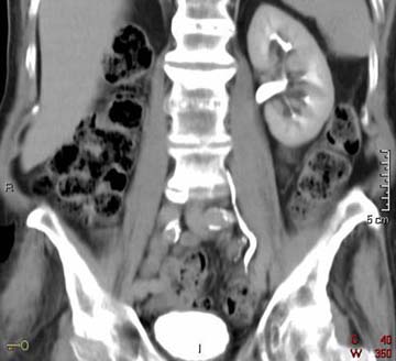
CT Scan showing right sided unilateral renal agenesis, available at: http://www.med-ed.virginia.edu/courses/rad/gu/embryology/agenesis.html
b. Bilateral Renal Agenesis: This condition is relatively uncommon and occurs mostly in males. It is characterised by olighydraminos (small amount of amniotic fluid), which causes compression of the fetus and leads to Potter syndrome (abnormal facial appearance, wrinkly skin and deformed limbs). As the condition is incompatible with life, the infants are usually stillborn or die shortly after birth.
Ectopic Kidney
It usually occurs when one or both kidneys fail to ascend and therefore remain abnormally present in the pelvic or lower abdominal area for example pelvic kidney.
Right sided ectopic kidney, available at: http://mamchenkov.net/wordpress/2005/04/14/ectopic-kidneys/
Renal Fusion
The most common type of renal fusion is the horseshoe kidney. In this anomaly the inferior poles of the kidney fuse across the midline. These kidneys fail to ascend because the fused kidneys are trapped behind the inferior mesenteric artery. The hilum lies ventrally because kidney rotation is also arrested. Although the function of kidneys is preserved, but it may sometime cause urinary tract obstruction due to impingement on the ureters and may lead to UTIs and pyelonephritis.
Horseshoe Kidney, available at: http://www.stanford.edu/dept/radiology/radiologysite/site392.html
Uretropelvic Duplication
It occurs when there is a duplication of both the ureter and renal pelvis (most commonly occurring) and also if there is an excess kidney present (very rare), termed as supernumerary kidney. These duplications occur due to division of metanephric diverticulum. Incomplete division leads to a divided kidney with a bifid ureter. Complete division results in a double kidney with bifid or separate ureters. If there is a formation of more than one metanephric diverticulum, it leads to an extra kidney being formed with its own ureter.
Fused supernumerary kidney, available at: http://radiology.casereports.net/index.php/rcr/article/viewArticle/552 |
Duplex kidney showing two ureters,available at: http://www.chop.edu/healthinfo/duplex-kidney.html |
Ectopic Ureter
When the ureteric bud fails to separate from the mesonephric duct, it results in the opening of the ureter distal to the normal position of ureters opening in the bladder. The most common ectopic opening is the lateral, and is termed as lateral ureteral ectopia.